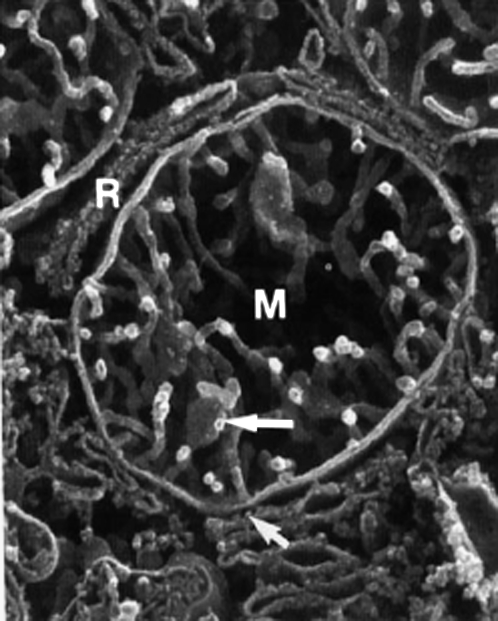
FIGURE 1.
High resolution scanning electron microscopy micrograph of human liver mitochondrion (M), in close proximity to rough endoplasmic reticulum (R). The cristae are tubular, several hundred nanometers long and ∼30 nm in diameter, × 40,500. Reprinted from Lea et al. (11) with permission of Wiley-Liss. In living cells, mitochondria are usually tubular (1–2 μm long and 0.1–0.5 μm large) and interact with other cellular components, especially with the cytoskeleton and endoplasmic reticulum (59,60).

