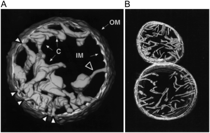
FIGURE 2.
Electron microscopy tomography images of mitochondria. Normally functioning mitochondria are going from (A) condensed to (B) orthodox state morphology, back and forth, depending on their respiratory rate. Mitochondria carrying out maximum respiratory rate (in excess ADP and respiratory substrate) present condensed (matrix contracted) morphology (A). Their cristae are swollen cisterns or sacs connected to the peripheral part to the IM by narrow tubular segments (cristae junctions) (A, large white arrow). The cristae in orthodox (matrix expanded) morphology are narrow, flattened, or almost tubular (b). OM diameter: (A) 1.5 μm; (B) lower, 1.2 μm. (A) Reprinted from Mannella et al. (13), with permission of IOS Press. (B) Reprinted from Mannella et al. (12) with permission of Wiley-Liss.

