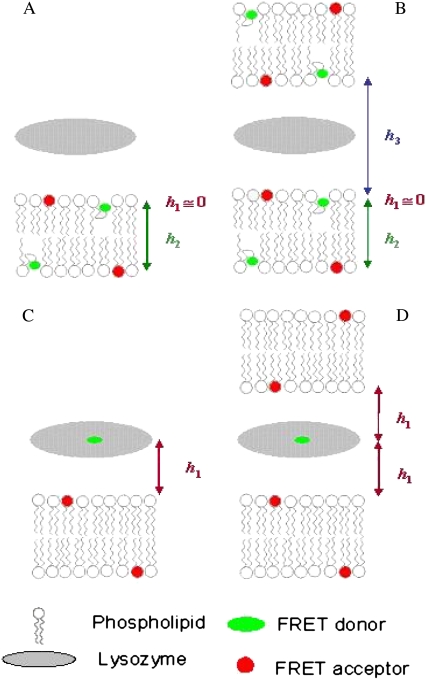
FIGURE 1.
Schematic representations of the FRET models used for both experimental setups explored in this work: FRET between two membrane probes (A and B), and where donors are located in the protein and acceptor is a membrane probe (C and D). A and C illustrate topological models for protein interaction with a single lipid bilayer, whereas B and D describe a multibilayer arrangement with protein molecules sandwiched between adjacent bilayers. Only two bilayers are depicted, because FRET to further acceptor planes is negligible. h1, h2, and h3 are the distances between planes of donors and acceptors taken into account in the FRET models.

