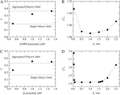
FIGURE 5.
(A) Acceptor numerical density, C, recovered from global analysis of fluorescence decays of FRET donor A488-lysozyme (D/P = 0.44) in the absence and presence of FRET acceptor Rh-PE (1 Rh-PE:400 total lipid) in 4:1 POPC/POPS vesicles (total lipid 215 μM). Experimental values (circles) are compared with theoretical expectations for single-bilayer (Fig. 1 C) and multibilayer (Fig. 1 D) models (dashed lines). (B) Global χ2 for the A488-lysozyme/Rh-PE pair samples, with [lysozyme] = 1.5 μM, as a function of the donor-acceptor interplanar distance (h1). The dashed lines are mere guides to the eye. (C and D) Same as A and B, but for the FRET pair lysozyme/DPH (1 DPH:200 total lipid).

