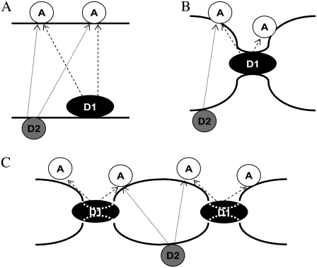
FIGURE 7.
Schematic representation of how the FRET pairs A488-lysozyme/Rh-PE (D1/A) and BODIPY-PC/Rh-PE (D2/A) probe differently the lamellar repeat distance in a pinched bilayer arrangement, as verified from the experimental data. (A) Low protein concentration (≤0.5 μM in the studied system). The protein binds to one vesicle without bridging adjacent bilayers. Repeat distances are high, and the recovered acceptor concentration is close to the single-bilayer expectation. (B) Intermediate protein concentrations (between 0.5 and 1.5 μM). Bridging occurs at the protein “pinches”. They are already reported by the protein donor D1 (because of its location at the pinches) but not by the derivatized lipid donor D2 (preferentially located away from these regions). (C) High protein concentrations (≥2.0 μM in the studied system). Bridging at the pinches is widespread and even the interbilayer distance sensed by FRET from D2 to A is reduced, though not as much as that reported by FRET from D1, for which a doubled acceptor concentration is sensed. D1 also inserts into the bilayer to some extent. Each thick solid line represents a lipid/water interface of a different bilayer, whereas molecules capable of undergoing FRET are united by dashed and dotted lines for the D1/A and D2/A pairs, respectively.

