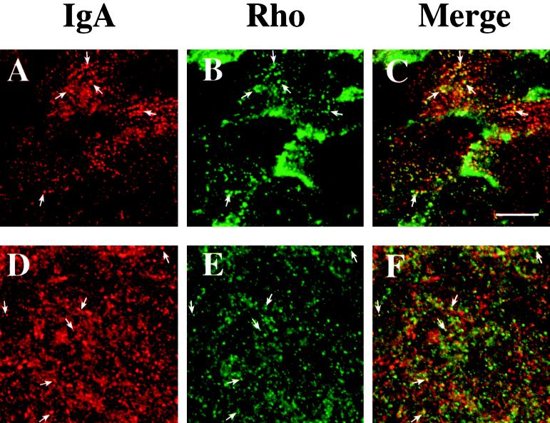
Figure 8.
Distribution of basolaterally internalized IgA and endogenous RhoA or myc-tagged RhoAV14. RhoAV14 cells were grown in the presence of 5 pg/ml DC (A–C) or 20 ng/ml DC (D–F). IgA was internalized from the basolateral pole of the cell for 10 min at 37°C, and cell surface ligand was removed by chasing in ligand-free medium for 5 min at 37°C (A–C) or by trypsin treatment at 4°C (D–F). The cells were fixed with paraformaldehyde, incubated with rabbit anti-IgA antibody (A–F) and a myc tag–specific mAb (A–C) or a monoclonal anti-RhoA antibody (D–F), and then reacted with goat anti-rabbit Cy5 and goat anti-mouse FITC secondary antibodies. Panel A is identical to panel H in Figure 7, and panel B is identical to panel M in Figure 1. In the latter case, the contrast was enhanced and the image brightened to demonstrate colocalization of myc-tagged RhoAV14 and IgA. Examples of IgA and endogenous RhoA or myc-tagged RhoAV14 colocalization are marked with arrows. Bar, 10 μm.

