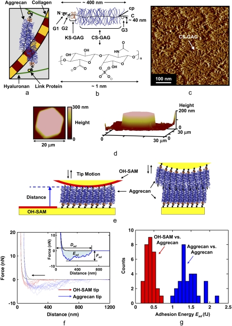
FIGURE 1.
Overview of the cartilage aggrecan macromolecule and experimental setup, and results of aggrecan self-adhesion. (a) Schematic of major load-bearing constituents of the ECM, including the type II collagen network and proteoglycan (aggrecan). Aggrecan macromolecules are attached to hyaluronan and stabilized via link protein. (b) Schematic of the cartilage aggrecan cylindrical brush-like structure (contour length, Lcontour ∼400 nm, molecular mass ∼3 MDa) (5), which is composed of a core protein backbone (cp) containing three globular domains (G1, G2, G3), grafted CS-GAG chains (Lcontour ∼40 nm, shown as C4S-GAG) with interchain spacing of ∼2–4 nm along the cp, and keratan sulfate GAG chains; N = N-terminal, C = C-terminal. (c) Tapping-mode AFM height image of two-dimensional closely packed fetal epiphyseal aggrecan on an atomically flat mica surface at a density that is ∼40× less than its physiological concentration in cartilage (adapted from Ng et al. (5), height scale <1 nm). (d) Contact-mode AFM two-dimensional (left, in 0.001 M NaCl, pH ∼5.6) and three-dimensional (right, in 0.1 M NaCl, pH ∼5.6) height image of a microcontact printed aggrecan-functionalized (inside the hexagonal pattern) and OH-SAM-functionalized (outside the hexagonal pattern) substrate imaged with an OH-SAM colloidal tip at 3 nN normal force (adapted from Dean et al. (11)). This image reflects a sample with the same aggrecan packing density as the nanomechanical experiments carried out in this study. (e) Schematics of colloidal force spectroscopy experiments reported in this work depicting the interactions between a functionalized planar substrate with end-grafted aggrecan and a hydroxyl-terminated monolayer (OH-SAM)-functionalized probe tip or an aggrecan end-grafted colloidal probe tip. (f) Comparison of colloidal force spectroscopy force-distance curves obtained via OH-SAM- and aggrecan-functionalized colloidal probe tips on aggrecan end-grafted planar substrates (1.0 M NaCl aqueous solution, surface dwell time t = 30 s, maximum compressive force, Fmax ≈ 45 nN, z-piezo displacement rate, z = 4 μm/s). Data for different experiments carried out at 10 different locations are shown for each probe tip. (Inset) Definition of the adhesive interaction distance, Dad, the maximum adhesion force, Fad, and the adhesion energy, Ead, for each pair of approach-retract force-distance curves. Statistically significant differences were observed for Fad of OH-SAM compared to aggrecan-functionalized probe tips (ANOVA, p < 0.001). (g) Histogram of Ead obtained via OH-SAM and aggrecan-functionalized colloidal probe tips on aggrecan end-grafted planar substrates (1.0 M NaCl aqueous solution, surface dwell time, t = 30 s, Fmax ≈ 45 nN, z-piezo displacement rate, z = 4 μm/s).

