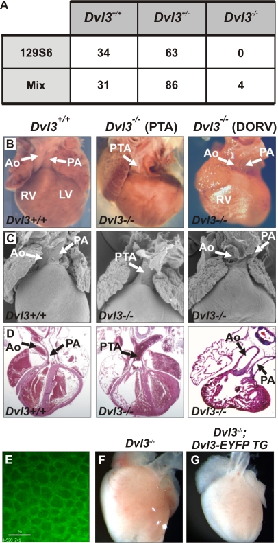
Figure 1. Cardiac defects of Dvl3 knockout mice.
The genotypes of weaning age pups from Dvl3 heterozygote crosses, in both an inbred 129S6 and mixed genetic background, were collected in order to calculate the frequency of survival of Dvl3 −/− mice (A). Cardiac abnormalities were observed in all Dvl3 −/− mutants examined in an inbred background (B–D). Whole mounts (B), scanning electron microscopy images (C) and hematoxylin and eosin stained sections (D) of P0 hearts of Dvl3 +/+ (left) and Dvl3 −/− hearts that displayed PTA (middle) and DORV (right). Aorta (Ao), pulmonary artery (PA), right ventricle (RV), left ventricle (LV). Deconvolution microscopy image showing EYFP-Dvl3 BAC transgene expression in the heart at E9.5. White bar = 20 µm (E). Addition of the Dvl3-EYFP transgene (G) rescues the abnormal cardiac phenotype of Dvl3 −/− mutants (F, heart displays PTA).

