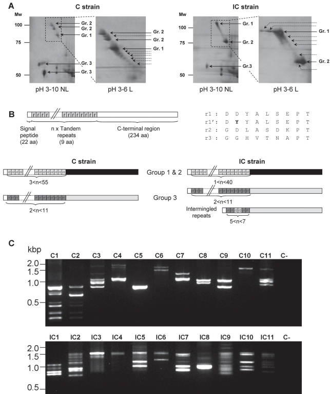
Figure 1. SmPoMuc polymorphism at the protein and transcript levels.
Positional differences between SmPoMuc from compatible (C) and incompatible (IC) strains on silver stained 2D-gels shown with a pH 3–10 non-linear (NL) gradient or a pH 3–6 linear (L) gradient (A). Positions of spots corresponding to SmPoMuc are indicated by arrows. Supplementary spots found in the present study using the pH 3–6 linear gradient are indicated by dotted arrows. (B) shows the precursor structure and polymorphism of SmPoMuc described in a previous study [18]. Three kinds of repeats were identified in SmPoMuc cDNAs (r1, r1' and r2); the fourth repeat r3 was only identified at the genomic level only in this study. (C) Agarose gel separation of RT-PCR amplicons obtained from 11 individual sporocysts (1–11) of both strains (compatible: C and incompatible: IC). Amplification was performed using consensus primers amplifying the complete coding sequence of all SmPoMuc. C-: negative control of amplification.

