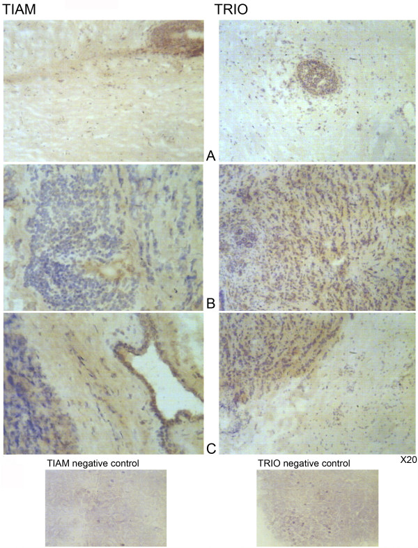
Figure 2.
Immunohistochemical staining of TIAM-1 and Trio in tumour and normal background breast tissue. Staining of TIAM-1 (left panel) and Trio (right panel) in normal background tissue (A) and in breast tumour tissue (B). Distribution of staining of TIAM-1 and Trio in tumour and surrounding normal tissue (C).

