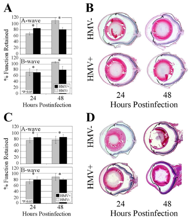
Figure 8. Histology and retinal function analysis of eyes injected with K. pneumoniae supernatant or metabolically inactive K. pneumoniae.

(A,B) Eyes were intravitreally injected with 0.5 μl of sterile-filtered supernatant of cultures of HMV+ or HMV− K. pneumoniae. Minimal retinal function loss (A) and inflammation (B) were observed with both groups at 24 and 48 h postinfection. (C,D) Eyes were intravitreally injected with 108 CFU equivalents of HMV+ or HMV− K. pneumoniae. Again, minimal retinal function loss (C) and inflammation (D) were observed with both groups at 24 and 48 h postinfection. Values represent the mean ± SEM for N≥4 eyes per time point and sections are representative of N=3 eyes per group.
