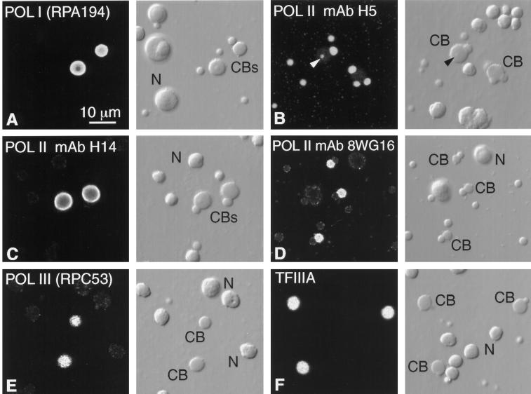
Figure 4.
Detection of RNA polymerases and TFIIIA by immunofluorescent staining. Each pair of panels shows the fluorescent image on the left and the corresponding DIC image on the right. CB, Cajal body; N, nucleolus. (A) Stain is brilliant in the matrix of Cajal bodies with a polyclonal antibody against the largest subunit of pol I, RPA194. For unknown reasons the nucleoli are very much weaker, although this antibody stains nucleoli well in HeLa cells. (B) Pol II stains strongly in B-snurposomes with mAb H5, which detects the CTD when serine 2 is phosphorylated (Patturajan et al., 1998), whereas the matrix of the Cajal bodies is only weakly stained. The arrowhead points to stained B-like inclusion in one Cajal body. (C) Pol II stains strongly in the matrix of Cajal bodies with mAb H14, which detects the CTD when serine 5 is phosphorylated (Patturajan et al., 1998), whereas the B-snurposomes are essentially unstained. (D) Pol II stains strongly in the matrix of Cajal bodies with mAb 8WG16, which recognizes the nonphosphorylated CTD (Patturajan et al., 1998). (E) Pol III stains strongly in the matrix of the Cajal bodies with a polyclonal antibody against the 53-kDa subunit, RPC53. (F) Pol III transcription factor TFIIIA is readily demonstrable in Cajal bodies with a polyclonal antibody.

