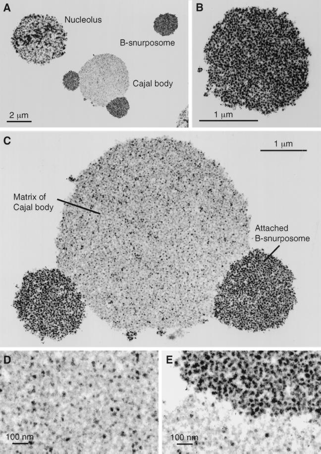
Figure 6.
Electron micrographs of thin sections through nuclear organelles, from a spread preparation of a Xenopus GV fixed in 4% glutaraldehyde and contrasted with OsO4, uranyl acetate, and lead citrate. (A) A nucleolus, a B-snurposome, and a Cajal body with two attached B-snurposomes. (B) A single B-snurposome composed of a relatively uniform aggregation of high-contrast 20- to 30-nm particles, which we call pol II transcriptosomes. (C) Cajal body from A at higher magnification showing that the matrix is heterogeneous in composition. The attached B-snurposomes are identical in appearance to free B-snurposomes. (D) At higher magnification the Cajal body matrix displays scattered 20- to 30-nm particles that look like pol II transcriptosomes, interspersed with numerous larger particles of lower contrast. (E) Boundary between the Cajal body matrix (below) and an attached B-snurposome (above) showing similar high-contrast particles in both compartments.

