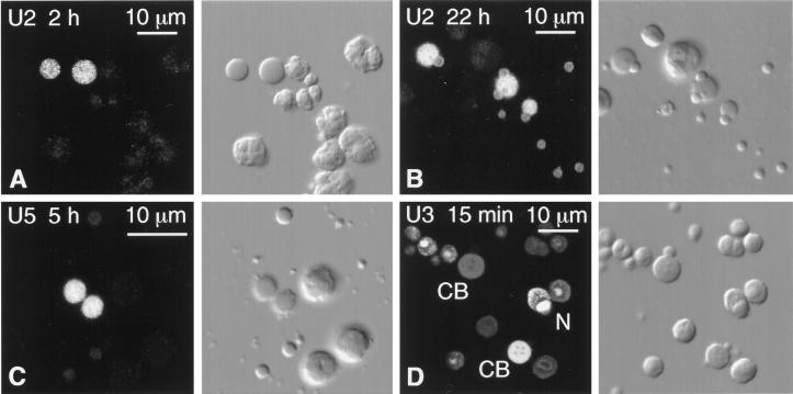
Figure 7.
Spread GV contents from oocytes injected with capped, fluorescein-labeled RNAs. The fluorescent signal was enhanced with goat anti-fluorescein labeled with Alexa 488. Each pair of panels shows the fluorescent image on the left and the corresponding DIC image on the right. CB, Cajal body; N, nucleolus. (A) Two hours after cytoplasmic injection of U2 snRNA, only the matrix of the Cajal bodies is labeled. (B) Twenty-two hours after injection of U2 snRNA, the matrix of the Cajal bodies is still labeled, but now the B-snurposomes are also labeled. (C) Five hours after nuclear injection of U5 snRNA, only the matrix of the Cajal bodies is significantly labeled. (D) Fifteen minutes after nuclear injection of U3 snoRNA, the matrix of Cajal bodies and the dense fibrillar region of the nucleoli are labeled. Individual Cajal bodies and nucleoli vary greatly in intensity.

