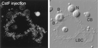
Figure 8.
Distribution of HA-tagged CstF77 in a GV spread, detected with an antibody against the HA tag. The fluorescent image is on the left; the corresponding DIC image is on the right. CB, Cajal body; N, nucleolus; B, B-snurposomes. The oocyte was injected 24 h previously with a transcript synthesized in vitro from a full-length cDNA clone of CstF77. The Cajal body is the most intensely stained structure, but label is detectable in the chromosome and the B-snurposomes.

