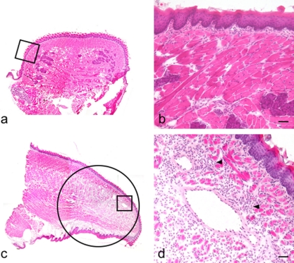
Fig. 2.
Induction of inflammatory reaction with TNF-α in the mouse tongue. H-E stained section (original magnification ×40). (a) Coronal section of the sham-injected radix linguae. (b) Region highlighted in a box in a. There is no inflammatory cell infiltration. (c) Sagittal section of the TNF-α-injected apex linguae. Inflammatory cell infiltration and vasodilation are observed in a circle. (d) Region highlighted in a box in c. Inflammatory cell infiltration and vasodilation are observed in the lamina propria mucosae and in the lingual tunica muscularis (arrowheads). Bar=100 µm.

