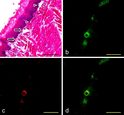
Fig. 4.
Expression of VCAM-1 on the mouse initial lymphatics of the radix linguae. (a) H-E stained section. (b) Immunostaining with anti-podoplanin of the section adjacent to a. Initial lymphatics expressing podoplanin in the lamina propria mucosae are visualized in green. (c) Immunostaining with anti-VCAM-1 of the section b. Vessels expressing VCAM-1 in the lamina propria mucosae are visualized in red. (d) Merged image of immunostaining with anti-podoplanin and anti-VCAM-1. There are initial lymphatics with the immunoreaction to anti-VCAM-1 (arrows) and also the vessels without the reaction (arrowheads). Bar=100 µm.

