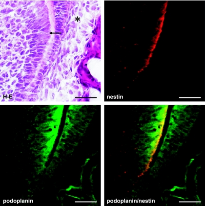
Fig. 4.
Podoplanin expression at the end of differentiating odontoblast layer. Figure shows the area of (c) in Figure 1 at a high magnification. Reaction products with anti-podoplanin antibody are observed on the differentiating odontoblasts at the junction with predentin (arrow), and weakly on the inner enamel epithelium at cell-cell contacts (asterisk). Bar=100 µm.

