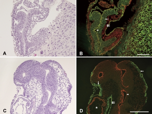
Fig. 1.
Hematoxylin and eosin staining of embryos at E9 (A) and E10 (C) stages in sagittal sections and double fluorescent immunohistochemistry for E-cadherin and N-cadherin in adjacent sections (B and D). Immunoreactivity of E-cadherin and N-cadherin are shown in green and red, respectively. In panel B, the single arrow and double arrow indicate the embryonic ectoderm and mesoderm, respectively. The arrowhead indicates the amniotic cavity. In panel D, the single arrows, double arrow, and an arrowhead indicate the surface ectoderm (epidermis), endoderm, and neuroectoderm, respectively. The heart primordial is indicated by an asterisk. Bars=100 µm.

