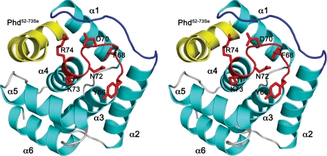
FIGURE 1.
Structure of Doc. A, stereo view of the DocH66Y-Phd52–73Se complex. Helices of DocH66Y are shown in cyan, and loop structures are shown in gray. The α-helices are labeled. The loop α3-α4 containing the conserved sequence motif HXFX(D/E)(A/G)N(K/G)R is highlighted in red, and its side chains are shown as sticks. Loop α1-α2 is highlighted in blue. The bound Phd52–73Se fragment is shown in yellow.

