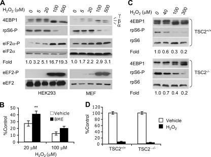
FIGURE 3.
Effects of H2O2 on mRNA translation and protein synthesis. A, HEK293 cells and immortalized wild-type mouse embryonic fibroblasts (MEFs) were exposed to 0-500 μm H2O2 for 1 h. Whole cell extracts were blotted for 4EBP1, phospho-rpS6, phospho-eIF2α, total eIF2α, phospho-eEF2, and total eEF2. Hypophosphorylated (α) and phosphorylated (β and γ) forms of 4EBP1 are indicated. Levels of total eIF2α and eEF2 proteins were examined for sample loading and protein stability using the same lysates run on separate gels. Changes in eIF2α phosphorylation (based on Image J analysis) compared with 0 μm H2O2 are shown. B, protein synthesis in H2O2-treated (1 h) MEFs with or without 2 h BME (100 μm) preconditioning. BME (100 μm) was present during the 1-h protein synthesis. **, p < 0.01. C, Western blotting for total 4EBP1 protein and rpS6 phospho-Ser235/236 in H2O2-treated (1 h) TSC2+/+ and TSC2-/- MEFs. Changes in rpS6 phosphorylation compared with 0 μm H2O2 are indicated. D, protein synthesis in TSC2+/+ and TSC2-/- MEFs treated with 100 μm H2O2 (1 h). [35 S]Methionine labeling was carried out in the presence of 100 μm H2O2. Base-line protein synthesis was similar in TSC2+/+ and TSC2-/- cells.

