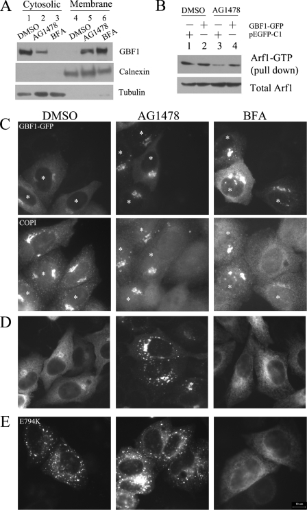
FIGURE 6.
The effect of AG1478 on GBF1. A, H4 cells were treated with DMSO (lanes 1 and 4), 14 μm AG1478 (lanes 2 and 5), 5 μm BFA (lanes 3 and 6) for 1 h, homogenized, and subjected to differential centrifugation. Equivalent amounts of the cytosolic and membrane fractions were resolved by SDS-PAGE and Western blotted with anti-GBF1, anti-calnexin, and anti-tubulin. B, H4 cells were transfected with ARF1-GFP together with GBF1-GFP or pEGFP-C1 for 24 h and then treated with DMSO, 14 μm AG1478 for 30 min. The cell lysates were analyzed by ARF1 pulldown assay. C, H4 cells were transfected transiently with GBF1-GFP plasmid for 24 h and treated with DMSO, 14 μm AG1478, or 1 μm BFA for 30 min. Cells expressing GBF1-GFP are indicated by white asterisks. The cells were stained with an antibody against β-COP. D, H4 cells were transfected transiently with GBF1-GFP plasmid for 24 h and treated with DMSO, 14 μm AG1478, or 5 μm BFA for 1 h. E, H4 cells were transfected transiently with E794K-GFP plasmid for 24 h and treated with DMSO, 14 μm AG1478, or 5 μm BFA for 1 h. Bar, 10 μm.

