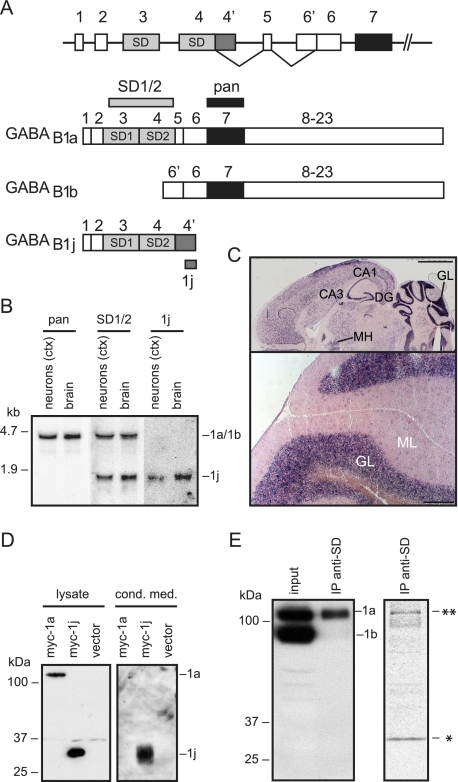
FIGURE 1.
Characterization of the GABAB1j isoform. A, schematic representation of the 5′ end of the GABAB1 gene indicating the exons encoding the GABAB1a, GABAB1b, and GABAB1j isoforms. GABAB1j results from an 870-bp extension of exon 4 at its 3′ end (exon 4′), generating an open reading frame of 687 nucleotides encompassing the two SDs. B, Northern blot analysis of GABAB1a and GABAB1j transcripts. Total RNA extracted from primary mouse cortical (ctx) neurons in culture or mouse brain was hybridized to the 32P-labeled probes indicated in A. The pan probe encodes part of the extracellular GABA binding domain and detects ∼4.5-kb GABAB1a and ∼4.1-kb GABAB1b transcripts (not resolved). The SD1/2 probe encodes the two SDs and detects GABAB1a and ∼1.6-kb GABAB1j transcripts. The 1j probe encodes 510 nucleotides at the 3′ end of exon 4′. C, in situ hybridization with the digoxigenin-labeled 1j probe. Top, horizontal section depicting the dorsal tier of the brain; bottom, high magnification of coronal section depicting lobules of the cerebellum. The locations of the CA1/3 field of hippocampus proper (CA1/3), dentate gyrus (DG), medial habenula (MH), and the granular layer (GL) and molecular layer (ML) of the cerebellum are indicated. Scale bars, 2 mm (top) and 200 μm (bottom). D, HEK293 cells expressing Myc-tagged GABAB1a (myc-1a) or GABAB1j (myc-1j) proteins. Conditioned medium (cond. med.) was subjected to immunoprecipitation with a rabbit anti-Myc antibody and analyzed in parallel with total cell lysate on Western blots using a mouse anti-Myc antibody. Membrane-bound GABAB1a protein was selectively detected in the cell lysate, whereas secreted GABAB1j protein was additionally detected in the cell-conditioned medium. E, left panel, the anti-SD monoclonal antibody 43H12 immunoprecipitates GABAB1a but not GABAB1b from mouse brain lysates. Immunoprecipitated GABAB1 protein (IP anti-SD) was analyzed in parallel with total brain lysate (input) on Western blots using a pan GABAB1 antibody (12). Right panel, the anti-SD monoclonal antibody 43H12 immunoprecipitates two proteins with a molecular mass corresponding to that of GABAB1a (**) and GABAB1j (*) from metabolically labeled cortical neurons. Radiolabeled proteins were revealed by autoradiography.

