FIGURE 2.
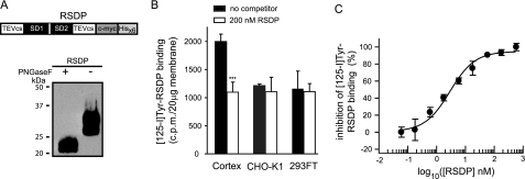
Specific binding sites for 125I-Tyr-RSDP in rat cortex synaptic membranes. A, expression of RSDP in Pichia pastoris. Top, a schematic representation of RSDP containing the two SDs flanked by two tobacco etch virus cleavage sites (TEVcs) and C-terminal c-Myc and polyhistidine (Hisx6) tags. Bottom, recombinant protein identified on Western blots using anti-His6 antibodies. RSDP is N-glycosylated as indicated by the shift from ∼29 kDa to the calculated molecular mass of ∼23 kDa after peptide N-glycosidase F (PNGaseF) treatment. RSDP is stable at 37 °C for at least 7 days (data not shown). B, 125I-Tyr-RSDP (0.5 nm) binding to 20 μg of membranes from cortex, CHO-K1, and HEK293FT cells, in the absence or presence of 200 nm unlabeled RSDP protein. Data are means ± S.D. from three independent experiments. C, inhibition of 125I-Tyr-RSDP (0.5 nm) binding to 40 μg of cortical membranes by different concentrations of unlabeled RSDP. The inhibition curve was calculated using nonlinear regression. Data points are means ± S.E. from three independent experiments.

