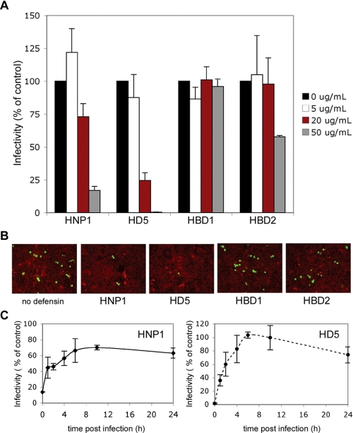
FIGURE 1.
HNP1, HD5, and HBD2 block BKV infection of Vero cells. A, Vero cells plated at subconfluent density were infected with BKV (m.o.i. 4) in 2% FBS containing MEM at 37 °C for 1 h. Indicated concentrations of defensins were present during infection. Cells were washed to remove unbound virus and 5% FBS containing MEM with the same concentrations of defensins was added to the cells and remained present for the duration of the experiment. Cells were fixed at 72 h and infection was determined by scoring the number of cells expressing the viral protein V-antigen (V-Ag). The graph represents the average of three experiments each of which have been normalized to infected cells without defensin treatment. Error bars represent the standard deviation. B, representative images of BKV-infected Vero cells are shown (magnification ×100). V-Ag-expressing cells are in green and Evans blue cytoplasmic labeling in red. C, Vero cells were infected with purified BKV (m.o.i. 4) in 2% FBS containing MEM at 37 °C for 1 h. Unbound virus was removed by washing and complete media added to cells. At the indicated times, 50 μg/ml HNP1 or HD5 was added to culture media. At the 0-h time point, defensins were added during and also directly following infection. Cells were stained and scored for V-Ag expression at 72 h post-infection. The graph shows the average of three experiments, each of which have been normalized to infected cells without defensin treatment. Error bars represent the standard deviation.

