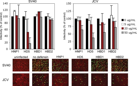
FIGURE 6.
HD5 inhibits SV40 and JCV infection. Subconfluent Vero cells or SVG-A were infected with SV40 (m.o.i. 2) or JCV (m.o.i. 5), respectively, in 2% FBS containing MEM at 37 °C for 1 h in the absence or presence of defensins at the indicated concentrations. After infection, cells were washed twice with MEM to remove unbound virus, and complete media containing defensins was added to cells for the duration of the incubation. Cells were fixed at 48 (Vero) and 72 h (SVG-A) post-infection and stained for V-Ag. Approximately 8,000 cells were screened for V-Ag expression. The graph shows the average of three experiments each of which has been normalized to infected cells without defensin treatment. Error bars represent the standard deviation. The bottom panel shows representative images of infected cells at the 50 μg/ml defensin concentration (magnification ×100). V-Ag-expressing cells are in green and Evans blue cytoplasmic labeling in red.

