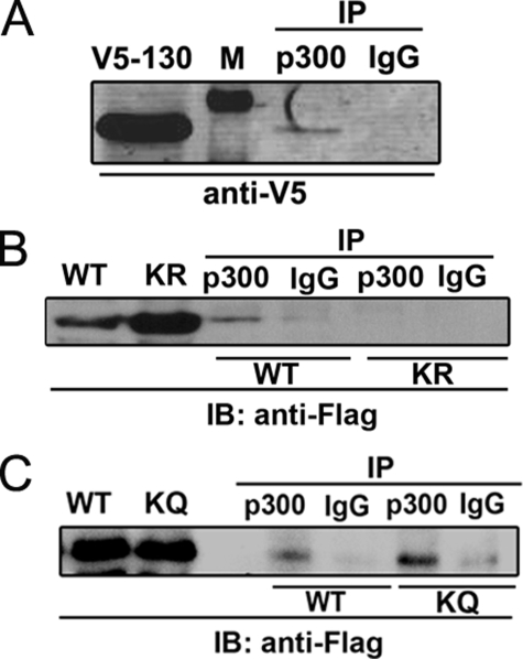
FIGURE 4.
Interaction between the STAT3 NH2-terminal domain and p300. A, HepG2 cells were cotransfected with pEF6-V5-STAT3 (aa 1–130) with pCMVβ p300. Cells were treated with IL-6 (10 ng/ml) for 30 min before NE was prepared. 2 mg of NE was immunoprecipitated by anti-p300 Ab. p300-bound V5-STAT3 (aa 1–130) was detected by anti-V5 Ab in Western immunoblot. Lane 1 is lysate showing the expression of V5-STAT3 (aa 1–130). Lane 2 is protein standard (indicated by “M” in A). B, HepG2 cells were cotransfected with either pECFP-FLAG-STAT3-WT (aa 1–124) or pECFP-FLAG-STAT3-K49R/K87R mutant (aa 1–124) together with pCMVβ p300. NE was immunoprecipitated with anti-p300 Ab followed by Western immunoblot (IB) with anti-FLAG Ab. Lane 1 and lane 2 show FLAG-STAT3-WT (aa 1–124) and FLAG-STAT3-K49R/K87R (aa 1–124) expression in nuclear lysate. Lane 3 and lane 4 are immunoprecipitates (IP) of FLAG-STAT3-WT (aa 1–124) with anti-p300 Ab (lane 3) or IgG (lane 4). Lane 5 and lane 6 are immunoprecipitates of FLAG-STAT3-K49R/K87R mutant (aa 1–124) with anti-p300 Ab (lane 5) or IgG (lane 6). C, HepG2 cells were cotransfected with either pECFP-FLAG-STAT3-WT (aa 1–124) or pECFP-FLAG-STAT3-K49Q/K87Q mutant (aa 1–124) (KQ) together with pCMVβ p300. Immunoprecipitation was performed as described in Fig. 1B. Lane 1 and lane 2 show FLAG-STAT3-WT (aa 1–124) and FLAG-STAT3-K49Q/K87Q (aa 1–124) expression in nuclear lysate. Lane 3 and lane 4 are immunoprecipitates of FLAG-STAT3-WT (aa 1–124) with anti-p300 Ab (lane 3) or IgG (lane 4). Lane 5 and lane 6 are immunoprecipitates of FLAG-STAT3-K49Q/K87Q mutant (aa 1–124) with anti-p300 Ab (lane 5) or IgG (lane 6).

