Figure 6.
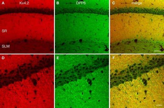
Enrichment of Kv4.2 and DPP6 in CA1 pyramidal cell dendrites. Confocal image of the CA1 field of the hippocampus in mouse double immunostained for Kv4.2 (A, pseudocolored red) and DPP6 (B, pseudocolored green) and the overlay of the two channels (C). Strong staining of the stratum radiatum (SR) was evident with both antibodies, however DPP6 antibodies produced stronger staining in the SLM than Kv4.2. (D–F) Higher magnification confocal image of CA1. Strong staining with both antibodies is observed in pyramidal cells' apical and basal dendrites, while pyramidal cell somata are only weakly stained. Scale bar: 50 μm (A–C); 20 μm (D–F).

