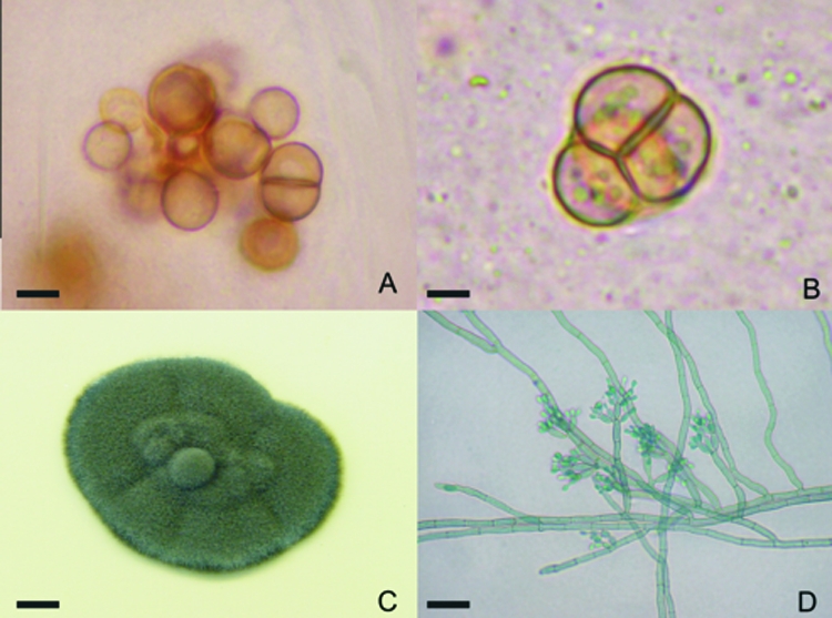
FIG. 1.

Fonsecaea pedrosoi sclerotic cells were obtained after scraping of chromoblastomycosis lesions. The cells gave rise to hyphae and conidia after in vitro culture. (A and B) Skin scrapings were collected and analyzed after clarification with 20% KOH, revealing well-defined, septated, sclerotic cells. (C) Greenish-black colonies grew from culture of this material on Mycosel. (D) Characteristic dematiaceous hyphae originating terminal cylindrical conidiophores with small subhyaline conidia were observed upon microculture. Bars, 4 μm (A), 3 μm (B), 0.5 cm (C), and 10 μm (D).
