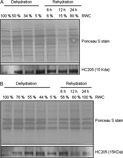
Fig. 9.
Western blot analysis of total protein extracts from X. humilis leaves (A) and roots (B) at different stages of desiccation and rehydration. The upper panels show the Ponceau S-stained nitrocellose membrane confirming equal loading and transfer of each experimental sample. The lower panel shows the Western blot result following the probing of these membranes with anti-Dsi-1VOC antibodies.

