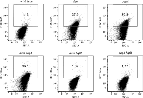
FIG. 6.
Flow cytometry analysis of std expression. Rabbit anti-StdA antiserum and FITC-conjugated goat anti-rabbit IgG were used for the detection of StdA antigen (y axes). Propidium iodide was used for the detection of DNA (x axes).The gate for the detection of StdA expression was set such that cells of the wild type (ATCC 14028) were considered positive for expressing StdA antigen when their FITC fluorescence intensity exceeded that of all but a small fraction (<2%) of the control population of the stdA mutant (not shown). The strains used were as follows: wild type (ATCC 14028), dam (SV4536), seqA (SV4752), dam seqA (SV4783), dam hdfR (SV5638), and seqA hdfR (SV5637). SSC-A, side scatter area.

