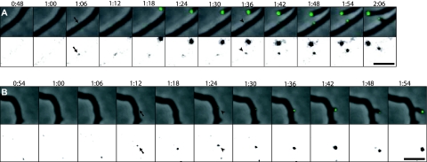
FIG. 1.
DivIVA marks sites of hyphal branching. Hyphae of strain K112 expressing divIVA-egfp were grown on agarose pads, and images were captured every 6 min. Time-lapse series of representative branching events are shown as overlays of fluorescence signal (green) on the phase contrast images (top) and as the fluorescence channel with the gray scale inverted and adjusted to clearly visualize the initially weak DivIVA-EGFP signals (bottom). The first time points when DivIVA-EGFP foci were detected at future branching sites are marked with arrows. The subsequent time points at which outgrowth of the branches can be seen are indicated by arrowheads. Time is shown in hours and minutes after starting time-lapse acquisition. Bars, 5 μm. The full time-lapse sequence from which the frames in panel A are derived is shown in Video S1 in the supplemental material.

