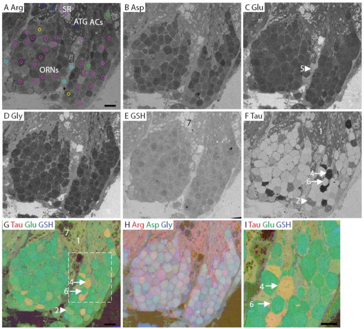
Figure 7.

Lobster olfactory (lateral) antennule. A: Arginine. The image is marked to indicate the main features of the antennule. The AOI mask is overlaid and color-coded (see legend in Fig. 3) by class: 1, blue; 2, yellow; 4, green; 5, cyan; 6, fuchsia; 7, purple (Class 3 see Fig. S4). B: Aspartate. C: Glutamate. D: Glycine. E: GSH. F: Taurine. G-H: RGB images of the lobster olfactory antennule. I: Higher magnification image of area within white dashed line in G. Numbers, arrows and arrowheads indicate the Class number. ACs, auxiliary cells; ATG, aesthetasc tegumental gland; SR, secretory rosette. Scale bars = 10 μm.
