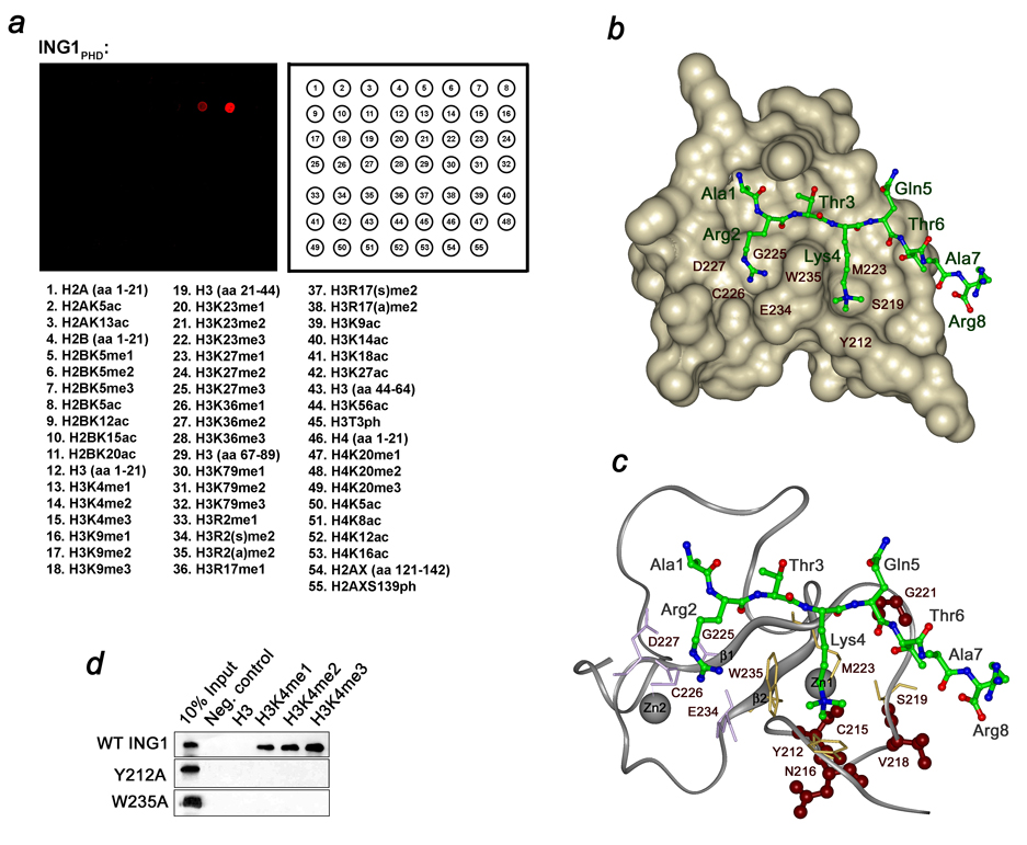
Figure 1. The ING1 PHD finger recognizes H3K4me3.
(a) Peptide microarrays containing the indicated histone peptides were probed with GST-ING1 PHD. Red spots indicate binding. me, methylation; ac, acetylation; ph, phosphorylation; s, symmetric; a, asymmetric. (b) Structure of the ING1 PHD finger in complex with the histone H3K4me3 peptide. The PHD finger is shown as solid surface. The histone peptide is depicted as ball-and-stick model with C, O and N atoms colored green, red and blue, respectively. (c) The PHD finger is shown as a ribbon with residues mutated in human cancers colored brown. (d) Interactions of the GST-fusion wild type and mutant ING1 PHD fingers with biotinylated histone peptides examined by western blot experiments.

