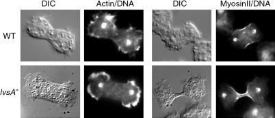
Figure 5.
Actin (left) and myosin (right) distribution in wild-type (top) and lvsA mutant (bottom) cells in attached conditions. Cells attached to coverslips were fixed and stained with DAPI to visualize their nuclei (DNA). Some cells were stained with rhodamine-labeled phalloidin to visualize actin filaments (Actin) or were transfected with an expression vector for the production of GFP–myosin II (MyosinII). The cells were then photographed by DIC microscopy (DIC) and fluorescence microscopy. The signal from the DAPI staining was merged with that from actin or myosin imaging. In attached conditions lvsA-mutant cells can organize their cytoskeleton like wild-type cells. Actin is found at the cortex of the cells predominantly at the poles, and myosin II is concentrated at the cleavage furrow. LvsA-mutant cells also display broad lamellipodia at the poles of the cells, but this does not seem to affect their ability to divide on a substrate.

