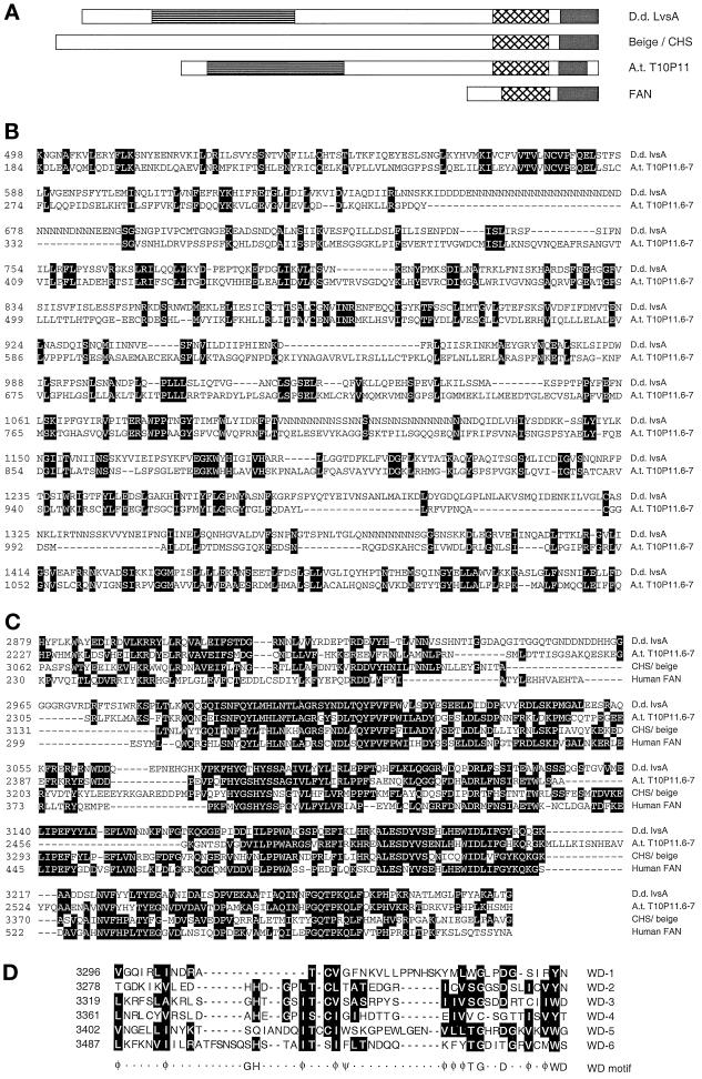
Figure 9.
The Dictyostelium lvsA protein contains domains similar to those found in beige and CHS, FAN, and a plant protein. (A) Diagram indicating the relative size of the mouse beige and human Chediak–Higashi syndrome proteins (Beige/CHS), the human FAN protein (FAN), and a hypothetical protein found in the Arabidopsis thaliana genome (T10P11.6–7, accession numbers 2262139 and 2262140 [A.t. T10P11]). The horizontal-hatched bars indicate the portions of LvsA (D. discoideum LvsA [D.d. LvsA]) that are similar to A.t. T10P11 (shown in B). The cross-hatched bars indicate the BEACH domain (shown in C). The black bars indicate the regions containing WD motifs (shown in D). (B) Region of homology between LvsA (accession number AF088979; D.d. lvsA) and the hypothetical A. thaliana protein T10P11.6–7 (A.t. T10P11.6–7). (C) Alignment of the BEACH domains from LvsA, human Chediak–Higashi syndrome protein (accession number U67615; CHS/beige), human FAN protein (accession number Q92636), and the hypothetical A. thaliana protein T10P11.6–7. (D) The WD motif region of LvsA. The six WD motifs from LvsA are aligned with each other. White letters on a black background indicate those positions at which at least three repeats have amino acids with the same chemical properties. Dashes are gaps inserted to align the sequences. The consensus WD motif based on the structure of Gβ (Sondek et al., 1996) is shown below the six LvsA repeats. Φ indicates hydrophobic residues; ψ indicates aromatic residues.

