
Fig. 1.
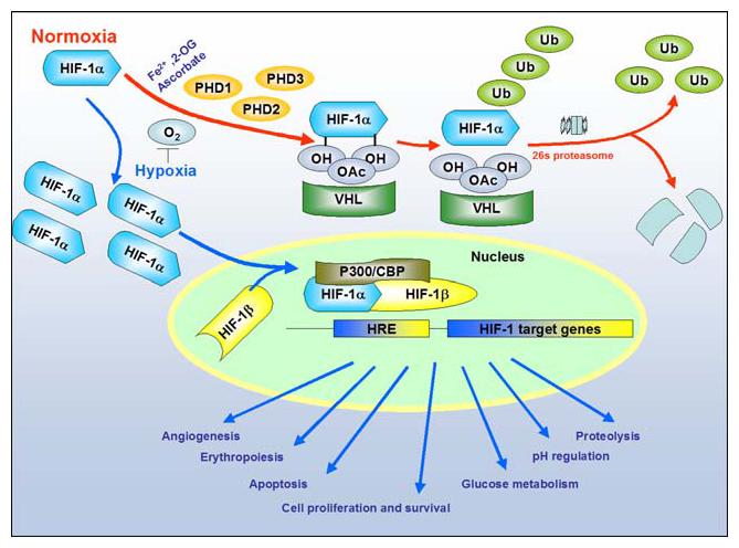
Cellular response to hypoxia: Levels of Hypoxia Inducible Factor-1 (HIF-1) are regulated by cellular oxygen by proline hydroxylation. The reaction is catalyzed by the enzymes prolyl 4-hydroxylases (PHD1:2 and 3). Under normoxia (red arrows), the intracellular level of HIF-1α is kept low by rapid ubiquitination and subsequent proteasomal degradation via recruitment of von Hippel-Lindau protein (pVHL), which depend on the hydroxylation of proline residues. In contrast, under hypoxia (blue arrows), both the intracellular level and the transcriptional activity of HIF-1α increase as a result of suppressed PHD activities. Consequently, HIF-1α forms a heterodimer with HIF-1β and changes the transcriptional rates of HIF-1-regulated genes under hypoxia (NOTE: For interpretation of the references to color in this figure legend, the reader is referred to the online version of this article)