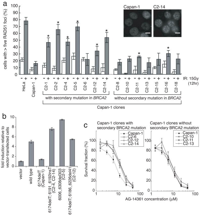
Figure 3. Functional analyses of the restored BRCA2 proteins.
a, Ionizing radiation (IR)-induced RAD51 foci formation is restored in most of BRCA2-restored Capan-1 clones. Indicated cells were irradiated (15 Gy) and fixed 12 hours after IR. Cells were immunostained with RAD51 antibody. Representative pictures of immunostained cells after IR are shown, together with quantification of the cells with at least five RAD51 foci before (−, white bars) and 12 hours after IR (+, grey bars) (mean values of at least three independent experiments ± SEM). Asterisks (*) indicate significant difference with irradiated parental Capan-1 cells (p<0.05, unpaired t test). Scale bar = 40 μm. b, Quantitation of HR induced by I-SceI in VC8-DR-GFP cells transiently transfected with wild-type and mutant forms of FLAG-tagged human BRCA2 cDNA. The proportion of GFP-positive cells for each construct relative to vector control is shown (mean ± SEM, n=3). c, BRCA2-restored Capan-1 clones are resistant to a PARP inhibitor. Capan-1, its clones with secondary BRCA2 mutation (C2-6, C2-12 and C2-14) and clones without secondary BRCA2 mutation (C2-10, C2-11 and C2-13) were treated with a PARP inhibitor (AG14361) at the indicated concentrations for 6 days, and survival fraction was measured by XTT assay (mean ± SEM, n=6).

