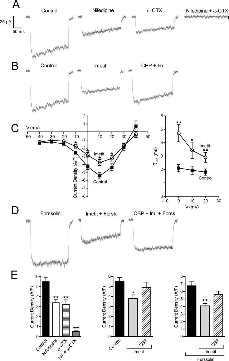
Figure 3.
Calcium current traces and peak current density recorded in NGF-differentiated PC12-H3 cells by stepping from the holding potential of −40 mV to the test potential of +10 mV. Panel A and E (Left): calcium current is inhibited by selective blockers of N- and L-type Ca2+ channels, i.e., ω-CTX (100 nM) and nifedipine (5 μM), both alone (n = 20) and in combination (n = 5). Panel B and E (Center) H3R activation with imetit (100 nM) attenuates calcium current, an effect prevented by the H3R antagonist clobenpropit (CBP; 50 nM). Panel C (Left): I-V curve in the absence and presence of imetit (100 nM) (means ± SEM; n = 20; *p < 0.05, from control by Student's t test). Panel C (Right): τact-voltage relationship. Imetit produced a slowing of current activation indicated by an increase in τact (means ± SEM; n = 12; *p < 0.05, **p < 0.01 from control by Student's t test). Panel D: Stimulation of adenylyl cyclase with forskolin (10 μM) increases calcium current. Incubation with imetit markedly reduces the forskolin-stimulated calcium current; CBP antagonizes this effect. Bars represent means (± SEM; n = 20; *p < 0.05, **p < 0.01 from control or forskolin by ANOVA followed by post hoc Dunnett's test).

