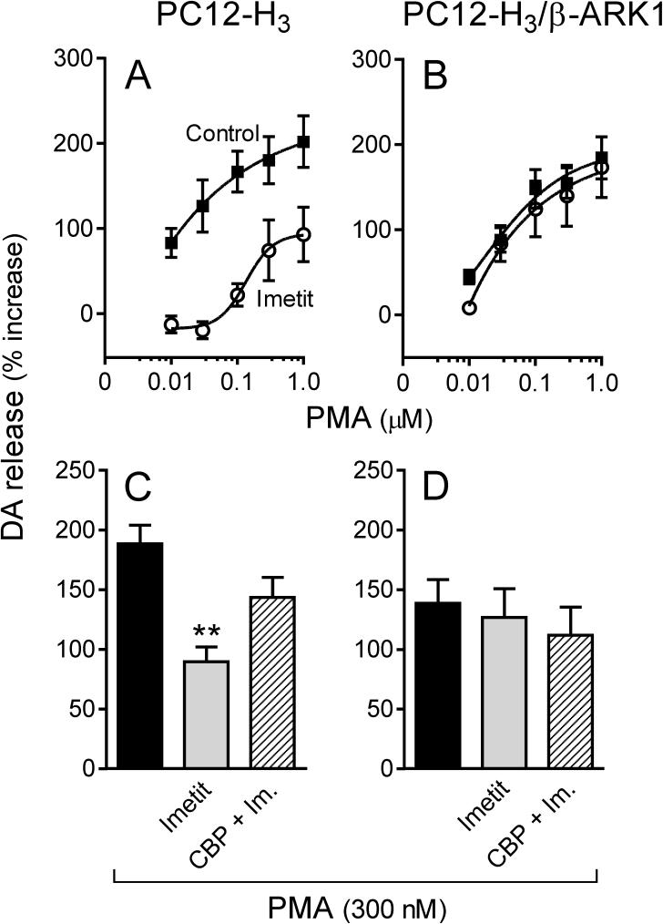
Figure 7.
Activation of H3R attenuates endogenous dopamine exocytosis elicited by PKC activation with PMA (300 nM) in PC12-H3 cells but not in PC12-H3/β-ARK1 cells. Panels A and B: concentration-response curves for PMA-induced dopamine release in PC12-H3 and PC12-H3/β-ARK1 cells, respectively. Activation of H3R with imetit (100 nM) significantly reduced PMA-induced dopamine release in PC12-H3 cells (A), an effect that was antagonized by CBP (50 nM; panel C). In PC12-H3/β-ARK1 cells (B), H3R activation with imetit (100 nM) failed to affect the PMA-induced dopamine release. Panels C and D represent dopamine release elicited by activation of PKC with PMA (300 nM) in the absence or presence of imetit (100 nM), either alone or in combination with clobenpropit (CBP, 50 nM). Bars are means ± SEM (n = 10 for C and n = 15 for D). Significantly different from PMA alone (**p < 0.01 by ANOVA followed by post hoc Dunnett's test). Dopamine release is expressed as percent increase above basal level.

