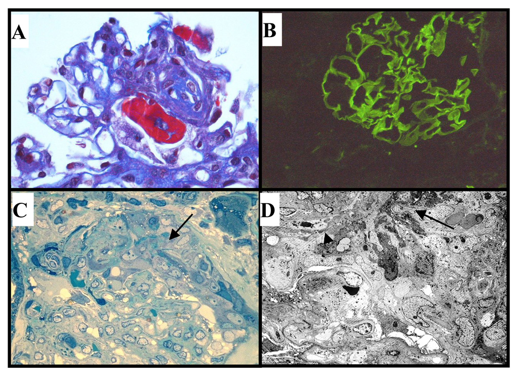
Figure 1. Diagnosis of IgA anti-GBM disease in the renal biopsy.
A. Light microscopy showing segmental fibrinoid necrosis (trichrome stain, x400). B. Direct immunofluorescence demonstrates strong (3+) linear capillary loop IgA (x200). C. Resin-embedded section showing active cellular crescent (arrow) composed of active cells with abundant cytoplasmic organelles and fibrinous exudate overlying compressed capillary tuft (toluidine blue, x400). D. Electron microscopy showing cellular crescent, focal fibrinoid damage (large arrow) and probable rupture of GBM (small arrow). Epithelial cell foot process fusion was present in relation to the crescent, but foot processes were largely intact in the other glomerulus (x800).

