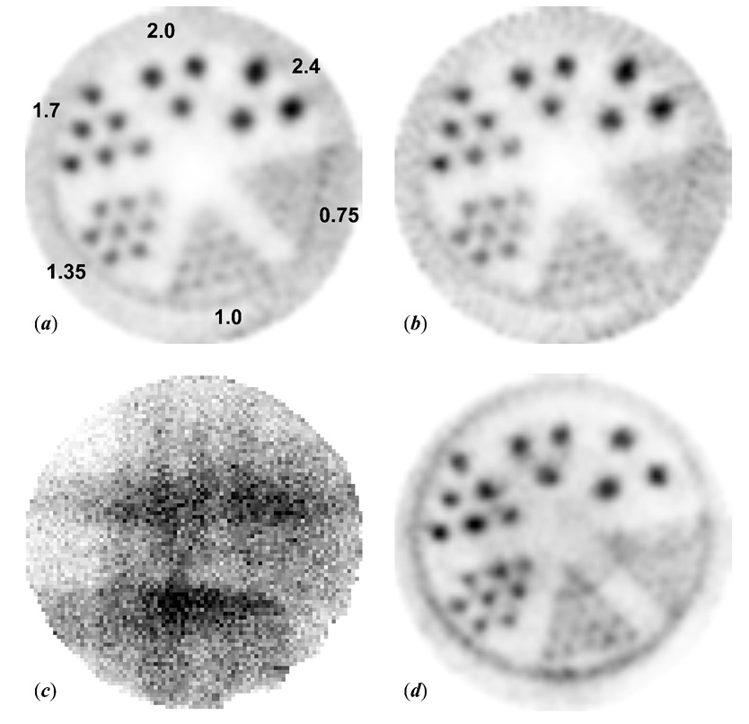
Fig. 8.
(a) A transaxial view of the hot-rod phantom using single-pinhole helical SPECT with 3° angular increments and 0.1 mm step increments along the AOR. Each of the 120 projections was 3 min/projection. The hot-rod phantom contained 10 MBq 125I with a concentration of 2 MBq/ml. The hot rod diameters of the six wedge-shaped regions in the phantom are 0.75, 1.0, 1.35, 1.7, 2.0, and 2.4 mm, respectively. (b) An image reconstructed from 60 slices out of those 120 projections to represent three-hour 1pHSPECT (c) One 3-min projection of the hot-rod phantom of a three-pinhole helical SPECT scan with the same imaging parameters as the 1pHSPECT scan. (d) A transverse image reconstructed from the 120 projections of the 3pHSPECT scan. Each reconstructed image presented here is 4.4 mm in thickness.

