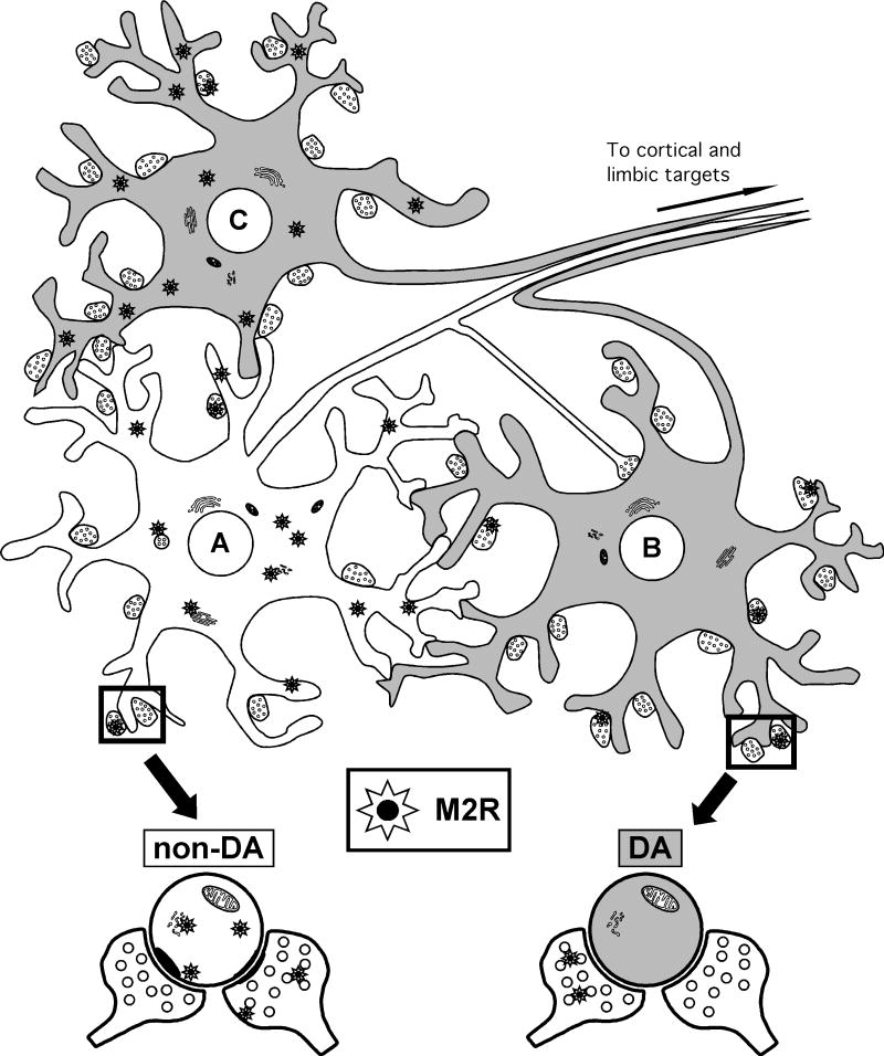
Figure 10.
Schematic diagram showing the distributions of M2 muscarinic receptors (stars) in neuronal profiles (somata, dendrites and terminals), which express (gray filling) or not (white filling) dopamine transporter (DAT) in the rat VTA. The M2 receptor is located mainly in non-DAT (presumably GABA-containing) neurons (A) receiving inputs from M2-labeled and/or unlabeled terminals. These contacts were mainly symmetric (inhibitory-type), but almost one-third were asymmetric (excitatory-type). The DAT-containing neurons (A) of the VTA, whose dendrites form multiple contacts with those of non-DAT neurons (B,C), usually do not express M2 receptor (B), but receive numerous contacts from M2-immunolabeled axon terminals. However, a minor proportion of DAT-containing neurons also express M2 receptor (C).

