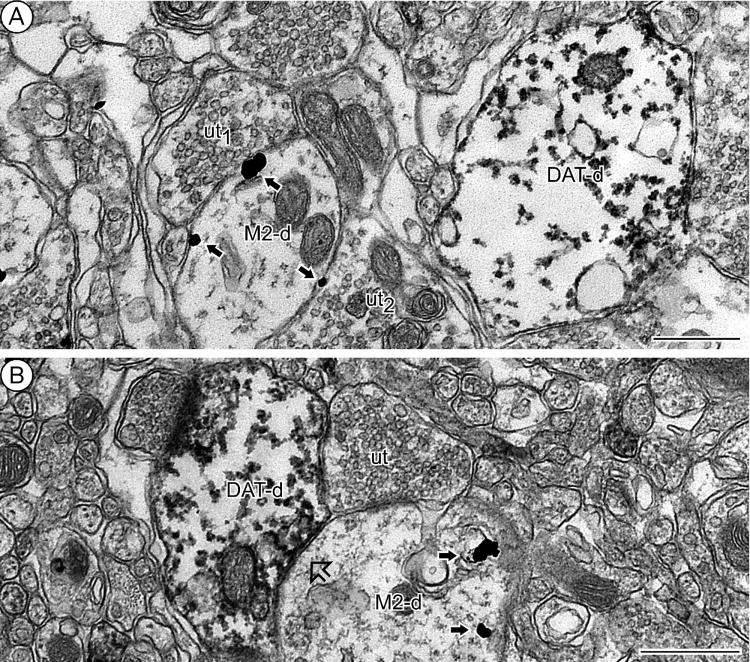
Figure 3.
Plasmalemmal M2 receptor distribution in non-dopaminergic dendrites. A: M2-immunogold particles (straight black arrows) are exclusively localized to the plasma membrane of a dendrite (M2-d) distinct from a nearby dendrite containing peroxidase labeling for DAT (DAT-d). The gold particles are located within and near contacts from unlabeled terminals (ut1-2). B: Unlabeled terminal (ut) establishes contacts with two differentially labeled and apposing (block arrow) dendrites; one is a M2-immunoreactive dendrite (M2-d) showing M2-immunogold particles (straight black arrows) and the other is a DAT-immunolabeled dendrite (DAT-d) containing DAT-immunoperoxidase reaction product. Scale bars = 0.5 μm.

