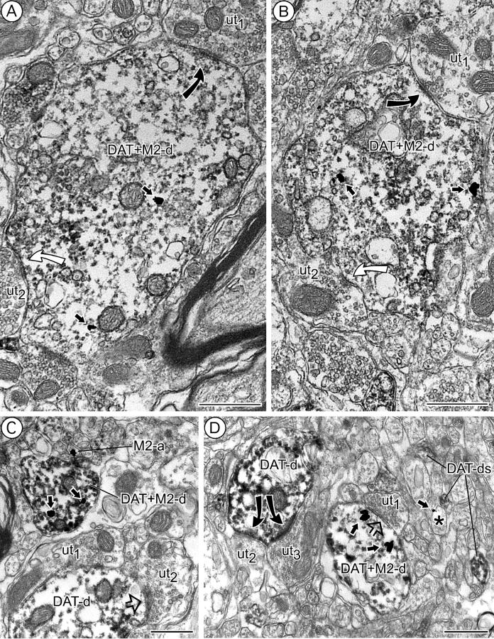
Figure 6.
Colocalization of M2 receptor and DAT in VTA dendrites. A: Immunoperoxidase reaction product for DAT is seen in a large dendrite (DAT+M2-d) showing sparse cytoplasmic M2-immunogold labeling. The dendrite receives both asymmetric (curved black arrow) and symmetric (curved white arrow) synapses from unlabeled terminals (ut1,2). B: Unlabeled terminals (ut1,2) establish symmetric (curved white arrow) or asymmetric (curved black arrow) synapses onto a dendrite (DAT+M2-d) containing light DAT-immunoperoxidase and M2-immunogold particles (straight arrows). C: A dually labeled small dendrite (DAT+M2-d) contains cytoplasmic M2-immunogold particles (straight arrows) and also intense immunoperoxidase reaction product for DAT. Another DAT-immunolabeled dendrite (DAT-d) contacting unlabeled axon terminals (ut1,2) is seen in close proximity. M2-a, M2-immunoreactive small unmyelinated axon. D: An unlabeled axon terminal (ut1) is apposed (open block arrow) to a dendrite (DAT+M2-d) showing immunoperoxidase reaction product for DAT as well as cytoplasmic M2-immunogold labeling (small black arrows). A nearby DAT-immunoreactive dendrite (DAT-d) receives convergent asymmetric inputs (curved black arrows) from unlabeled terminals (ut2,3). In the surrounding neuropil, there are additional DAT-immunoperoxidase labeled dendrites (DAT-ds), one of which apposes a small M2-immunogold labeled (straight arrow) profile presumed to be a dendrite (asterisk). Scale bars = 0.5 μm.

