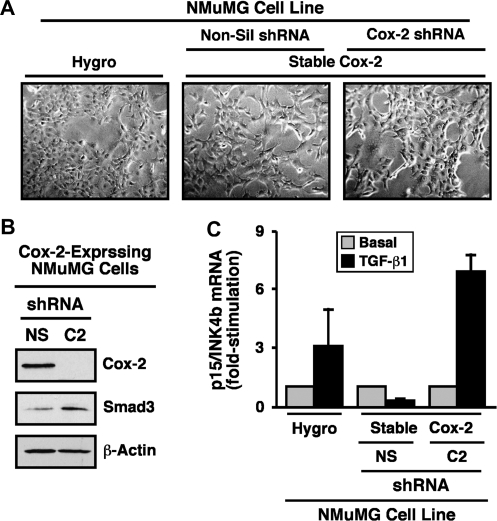
Fig. 6.
Cox-2 deficiency reverts fibroblastoid morphology and restores TGF-β coupling t Smad3 in NMuMG cells. (A) Cox-2-expressing NMuMG cells were subjected to lentiviral-mediated transduction of control (i.e. non-silencing) or Cox-2 shRNA as indicated and subsequently were selected for by resistance to puromycin. Afterward, bright field images of parental (i.e. Hygro), Cox-2-expressing (i.e. non-silencing, Non-sil) or Cox-2-deficient (i.e. Cox-2 shRNA) NMuMG cells were captured from a representative experiment that was performed three times with identical results. (B) Cox-2-expressing (i.e. non-silencing shRNA, NS) or Cox-2-deficient (i.e. Cox-2 shRNA, C2) NMuMG cells were harvested and immunoblotted with antibodies against Cox-2, Smad3 and β-actin as indicated. Images are from a representative experiments that was performed three times with identical results. (C) Parental (i.e. Hygro), Cox-2-expressing (i.e. non-silencing, NS) or Cox-2-deficient (i.e. Cox-2 shRNA, C2) NMuMG cells were incubated for 36 h in the absence or presence of TGF-β1 (5 ng/ml) as indicated. Afterward, total RNA was isolated and subjected to semiquantitative real-time PCR to monitor the expression of p15/INK4b. Data are the mean (±SE; n = 2) fold changes of plasminogen activator inhibitor-1 gene expression relative to unstimulated control cells (*P < 0.05; Student's t-test).

