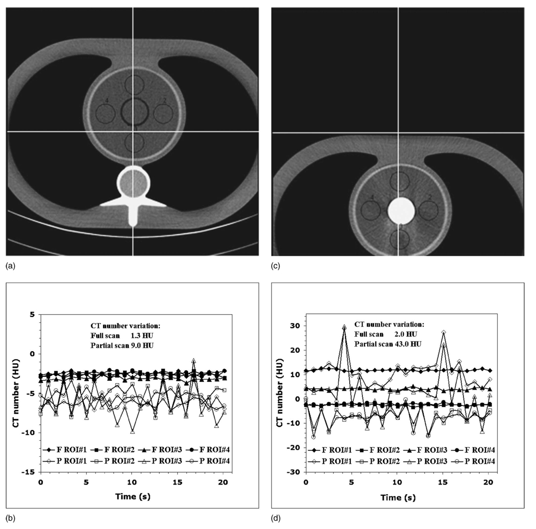
FIG. 6.
(a) The cardiac phantom with the water-filled syringe aligned at scanner isocenter. (b) The phantom geometry has enough anisotropy to cause mild to moderate partial scan artifacts, depending on the ROI location. (c) The cardiac phantom with the iodine-filled (2000 HU) syringe located 10 cm off the isocenter. (d) This geometry has large anisotropy, resulting in moderate to severe partial scan artifacts, depending on the ROI location.

