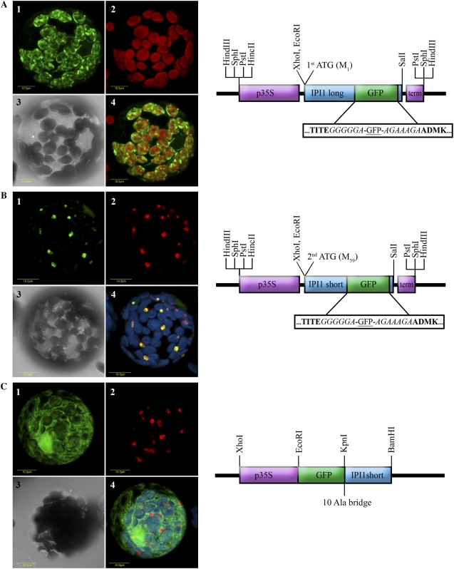
Figure 3.
Localization of Arabidopsis IPI1 versions by transient expression of IPI1-GFP fusions in tobacco protoplasts. A, The full-length ORF of IPI1 encoded by the long transcript (starting from the first Met codon) was fused internally with GFP as schematically shown and as described in the methods. The construct was transiently transformed into tobacco protoplasts, which were visualized using a laser scanning confocal microscope. A representative protoplast is visually presented as follows: section 1, green fluorescence corresponds to GFP; section 2, red fluorescence corresponds to chlorophyll; section 3, bright-field image; section 4, confocal image recorded simultaneously for green and red fluorescence (i.e. GFP and chlorophyll fluorescence overlaid). B, The ORF of IPI1 encoded by the short transcript (starting from the second Met codon) was fused internally with GFP as schematically shown and as described in the methods. The construct was transiently transformed into tobacco protoplasts, which were visualized using a laser scanning confocal microscope. A representative protoplast is visually presented as follows: section 1, green fluorescence corresponds to GFP; section 2, red fluorescence corresponds to peroxisome marker Cherry-PTS1 (Avisar et al., 2008); section 3, bright-field image; section 4, confocal image recorded simultaneously for GFP (green fluorescence), Cherry-PTS1 (red fluorescence), and chlorophyll fluorescence (coded blue, overlaid). C, The ORF of IPI1 encoded by the short transcript (starting from the second Met codon) was fused N terminally to GFP as schematically shown and as described in the methods. The construct was transiently transformed into tobacco protoplasts, which were visualized using a laser scanning confocal microscope. A representative protoplast is visually presented as follows: section 1, green fluorescence corresponds to GFP; section 2, red fluorescence corresponds to peroxisome marker Cherry-PTS1 (Avisar et al., 2008); section 3, bright-field image; section 4, confocal image recorded simultaneously for GFP (green fluorescence), Cherry-PTS1 (red fluorescence), and chlorophyll fluorescence (coded blue, overlaid). Schematic representation of the constructs are as follows: p35S (purple box) designates CaMV 35S promoter; term (purple box) designates CaMV 35S terminator; GFP (green box) designates the ORF of the green fluorescent protein; IPI1 long and IPI1 short designate the ORF of Arabidopsis IPI1 starting from the first or second Met codon, respectively (blue box). Boxed sequence blowup shows amino acid residues flanking the GFP sequence; residues in bold are Arabidopsis IPI1 sequences; residues in italic are protein spacer sequences. 10 Ala bridge designates a spacer of 10 Ala residues between the GFP and IPI1 sequences.

