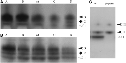
Figure 2.
PGM isoform patterns in potato and Arabidopsis as revealed by native PAGE followed by enzyme activity staining. Extracts from leaves (A) or growing tubers (B) of two overexpressing (lines A and B) and two underexpressing (lines C and D) transgenic lines from potato and from wild-type plants (wt) were separated by native PAGE. In A, 20 μg of protein was loaded in each lane; in B, 30 μg of tuber protein was applied per lane; in C, 20 μg of protein from leaves of Arabidopsis wild type (ecotype Columbia) or from the mutant lacking the plastidial PGM (p-pgm) was loaded in each lane. Following electrophoresis, the slab gel was stained for PGM activity. In A and B, the three PGM forms were designated 1 (having the highest mobility) to 3 (lowest mobility). Plastidial (white arrowheads) and cytosolic (black arrowheads) isozymes of PGM are marked. The location of form 2 (diamonds) remains to be clarified. In C, the white arrowhead marks the plastidial PGM isozyme (band I) and the two closed arrowheads (bands II and III) mark the two cPGM isozymes.

