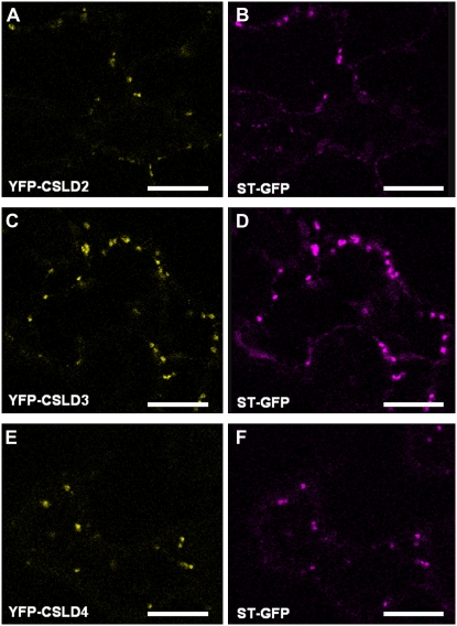
Figure 10.
Subcellular localization of CSLD1 and CSLD4. Fluorescently tagged versions of CSLD2 (A), CSLD3 (C), and CSLD4 (E) in which YFP was fused to the N termini were expressed in Nicotiana benthamiana leaves and visualized using confocal laser-scanning microscopy. The Golgi apparatus marker STtmd-GFP (ST-GFP) was also expressed in N. benthamiana leaves (B, D, and F). Bars = 15 μm.

